Biofilm Imaging & Structural Characterization
Biofilm Imaging & Structural Characterization provides direct, high-resolution insight into how microbial communities attach, organize, and mature on material surfaces. By visualizing biofilm architecture, thickness, viability, and adhesion, these studies reveal critical structural features that drive microbial persistence, resistance, and material interaction.
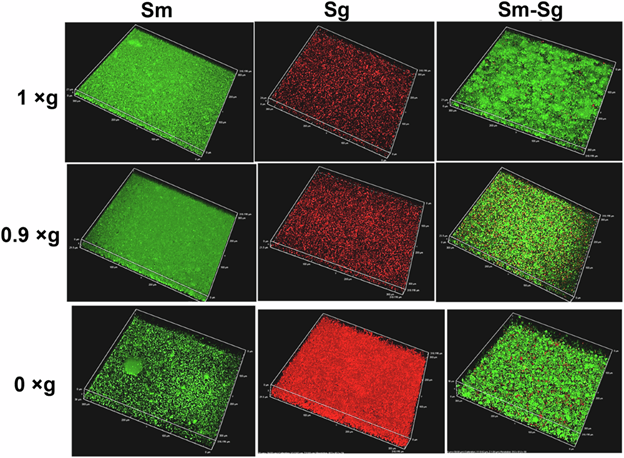
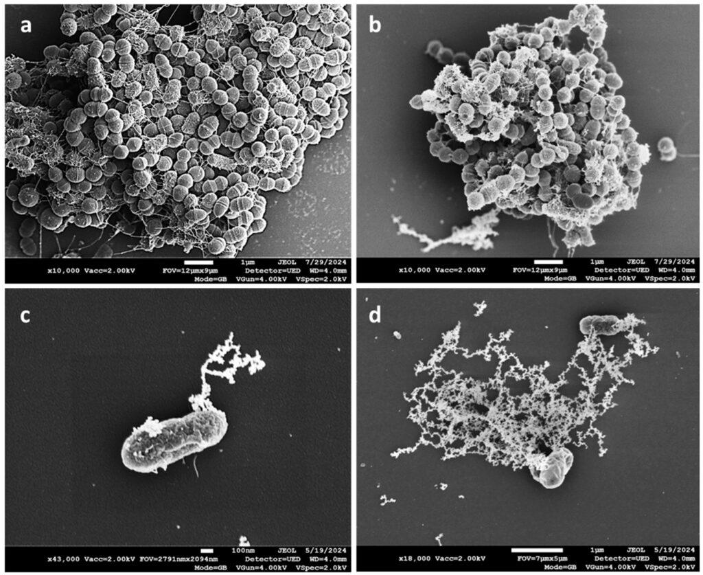
At Materials Metric, we integrate advanced imaging platforms, including SEM, CLSM, AFM, and fluorescence-based viability staining—to capture biofilm organization across micro- and nanoscale dimensions. This multi-modal approach enables precise evaluation of surface susceptibility, antimicrobial effectiveness, and biofilm disruption mechanisms, supporting materials development, device validation, and regulatory submissions where biofilm performance is critical.
Uses
• Evaluate treatment effects on biofilm morphology
• Study adhesion mechanisms and surface–biofilm interactions
• Compare materials, coatings, and antimicrobial strategies
• Characterize thickness, density, and biomass structure
Applications
• Medical device materials
• Dental and implant surfaces
• Polymers, hydrogels, composites
• Water and industrial surfaces
Sample Analysis Process
1. Sample Preparation
- Fixation, staining, dehydration, or cryo-prep
2. Imaging
- SEM for high-resolution structural imaging
- CLSM for 3D architecture and live/dead staining
- AFM for surface interaction mapping
3. Interpretation
- Biofilm thickness, density, and viability patterns
- Structural differences after treatment
Why Choose Materials Metric
We deliver publication-quality, high-resolution biofilm imaging with expert analysis that reveals biological mechanisms.
We offer:
• SEM, CLSM, AFM, and fluorescent imaging capabilities
• ISO 9001:2015 controlled preparation and imaging workflows
• Quantitative and qualitative structural assessments
• Deep expertise in surface science and microbial adhesion
Materials Metric provides unmatched clarity into biofilm behavior—critical for validating coating performance and microbial safety.
Related services